
Sleep apnoea can leave a person exhausted even after spending eight hours in bed, yet many people do not know why their breathing keeps stopping. The condition does not have one universal cause. In obstructive sleep apnoea, the throat repeatedly narrows or closes while the person sleeps. In central sleep apnoea, the airway may remain open, but the brain briefly fails to maintain the signals that control breathing. Body weight, jaw position, tongue size, tonsils, age, hormones, alcohol, smoking, medicines and certain health conditions can all influence the risk. Understanding sleep apnoea causes is important because the right treatment depends on the type of breathing problem and the factors contributing to it. Snoring or tiredness alone cannot confirm a diagnosis. A clinical assessment and sleep study may be needed to identify what is happening during sleep.
The most common cause of sleep apnoea is repeated narrowing or closure of the upper airway during sleep. This is known as obstructive sleep apnoea. Central sleep apnoea has a different cause: the brain temporarily fails to maintain the signals needed for normal breathing. Healthdirect explains that obstructive sleep apnoea involves partial or complete airway blockage, while central sleep apnoea results from pauses in the drive to breathe.
What Causes Sleep Apnoea?
The direct cause of sleep apnoea depends on which type a person has. Obstructive sleep apnoea, or OSA, is mainly a physical airway problem. The tongue and surrounding throat tissues restrict airflow even though the chest is still trying to breathe. Central sleep apnoea, or CSA, is a breathing-control problem in which breathing effort reduces or stops because of changes in signals from the brain.
| Type of sleep apnoea | What causes the breathing interruption? | Is the airway blocked? |
| Obstructive sleep apnoea | The upper airway repeatedly narrows or closes | Yes |
| Central sleep apnoea | The brain temporarily stops maintaining normal breathing signals | Usually no |
| Treatment-emergent central sleep apnoea | Central events appear during treatment of obstructive events | Not always |
| Mixed sleep apnoea | Obstructive and central events occur in the same person | Sometimes |
A direct cause is different from a risk factor. Upper-airway collapse is the direct cause of an obstructive event. Excess tissue around the neck, a large tongue or a lower jaw that sits further back can make that collapse more likely. Alcohol and back sleeping may worsen an existing airway problem. Conditions such as high blood pressure and type 2 diabetes are commonly associated with OSA, but they should not automatically be described as its original cause.
Why Does the Airway Close in Obstructive Sleep Apnoea?
The upper airway is a flexible breathing passage rather than a rigid pipe. Muscles around the tongue and throat help hold it open while a person is awake. Muscle tone naturally falls during sleep. Most people continue breathing normally, but an airway that is narrow or more likely to collapse may partially or completely close.
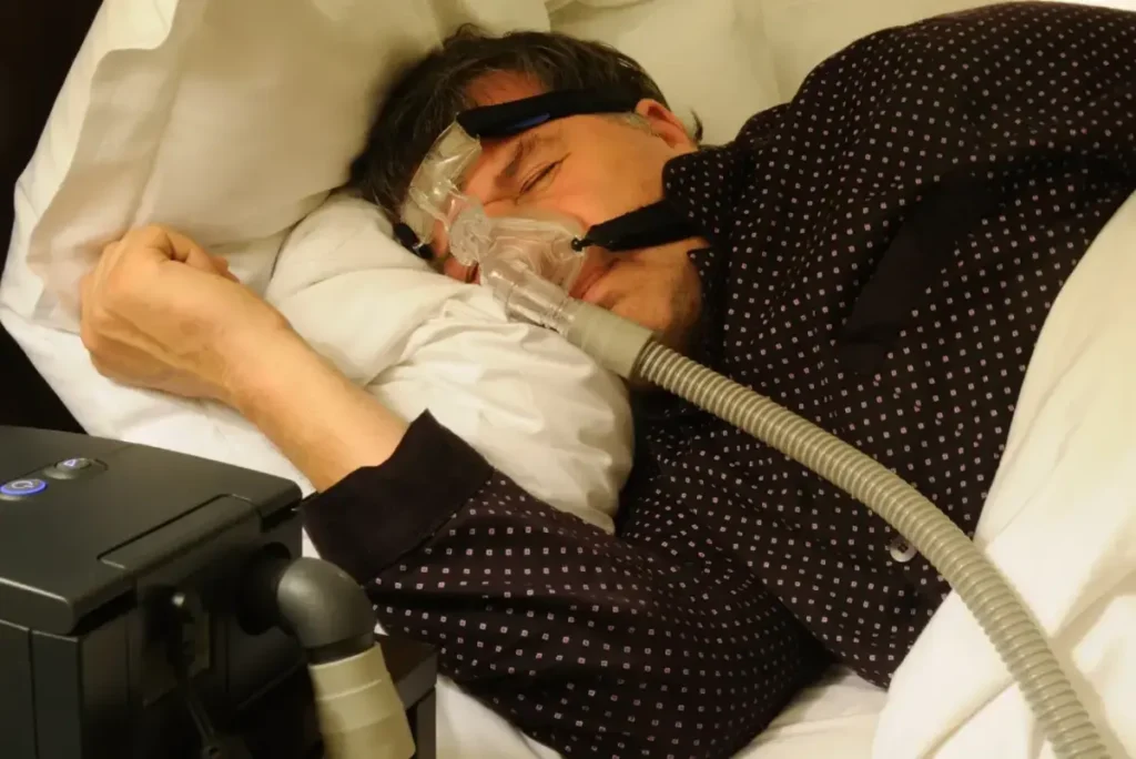
Throat Muscle Tone Decreases During Sleep
The muscles supporting the tongue, soft palate, uvula and throat walls become more relaxed during sleep. If enough space remains, air continues to move normally. If the breathing passage is already narrow, relaxed tissues may move inward and create resistance. Air passing through a narrowed space can make the soft tissues vibrate, producing snoring. The airway may then narrow further and reduce breathing. Snoring is common in OSA, but its loudness does not show how severe the obstruction is. Some people snore loudly without OSA, while others have significant breathing interruptions with less obvious snoring.
The Tongue and Soft Tissues Reduce Airway Space
The tongue base can move backwards when muscle support decreases. Tissue around the throat can also press inward. A lower jaw that sits further back may leave less room between the tongue and the back of the throat, which can increase the chance of obstruction. A partial reduction in airflow is called a hypopnoea. A more complete pause is called an apnoea. During an obstructive event, the chest and respiratory muscles may continue working, but little or no air moves through the narrowed airway. The person is attempting to breathe against the blockage.
The National Heart, Lung, and Blood Institute identifies the tongue, soft palate, neck tissue and upper-airway anatomy as factors that can block airflow during sleep.
The Brain Briefly Restores Breathing
When airflow falls, oxygen levels may decrease and carbon dioxide may change. Breathing effort can increase as the body tries to move air through the obstruction. The brain then causes a brief arousal, returning enough muscle tone to reopen the airway.
The sequence may look like this:
- The throat narrows or closes.
- Airflow reduces or stops.
- Breathing effort continues or increases.
- The brain briefly raises alertness.
- Throat muscle tone returns.
- The airway opens, sometimes with a gasp or snort.
- The person falls asleep again.
- The same cycle may repeat.
These arousals are often so brief that the person does not remember them. However, repeated interruptions can prevent deep, restorative sleep and lead to daytime tiredness, poor concentration and morning headaches. Healthdirect notes that OSA breathing pauses may happen many times during the night without the person being aware of them.
Why the Cycle Can Be Worse at Certain Times
Obstruction may become more likely during rapid eye movement, or REM, sleep because muscle tone is lower. Sleeping flat on the back can also allow the tongue and soft tissues to move further into the breathing passage. Alcohol, severe tiredness, nasal congestion and some sedating substances may make an existing airway problem worse. These factors usually expose or aggravate an underlying vulnerability rather than creating every case of OSA on their own.
What Increases the Risk of Obstructive Sleep Apnoea?
Obstructive sleep apnoea can affect people of different ages, body types and backgrounds. Some risk factors relate to body tissues and airway anatomy. Others involve hormones, medical conditions, family history or habits that influence muscle tone and breathing. Healthdirect identifies excess weight, male sex, post-menopausal status, a thick neck, a naturally narrow airway, a large tongue, enlarged tonsils or adenoids, age, family history, alcohol and smoking as recognised risk factors.
Body Weight and Tissue Around the Airway
Living with overweight or obesity can increase the amount of tissue around the neck, throat and tongue. This may reduce the airway’s resting size and make it easier for the walls to collapse during sleep. Weight is an important risk factor, but it is not the only explanation. A person at a healthy body weight may still have OSA because of jaw position, tongue size, tonsils, age, family history or another airway feature. Likewise, living with obesity does not automatically mean that a person has sleep apnoea.
Neck circumference can be useful during risk assessment because it may reflect tissue around the airway. However, no neck measurement can diagnose OSA on its own. Clinical history and sleep testing remain important.
Jaw, Tongue and Upper-Airway Anatomy
The relationship between the lower jaw, tongue and throat can influence the space available for breathing. A smaller lower jaw, or one that sits further back, can leave less room behind the tongue. During sleep, the tongue may then move backwards and restrict airflow.
Other anatomical factors can include:
- A relatively large tongue
- Enlarged tonsils or adenoids
- A naturally narrow throat
- A long or thick soft palate
- Craniofacial differences
- A narrow upper jaw
- Nasal obstruction
- Reduced space behind the tongue
No single dental feature proves that OSA is present. An oral and jaw examination can identify features that may affect treatment choices, but diagnosis generally requires medical assessment and sleep testing. This anatomical link is also why mandibular advancement splints may help selected patients. The appliance holds the lower jaw forward, which can increase the space behind the tongue. Oral appliance therapy is relevant to obstructive airway collapse, not central sleep apnoea.
Age, Sex, Menopause and Pregnancy
Sleep apnoea can develop at any age, but the risk generally increases through middle and older age. Changes in muscle tone and the distribution of tissue around the neck and tongue may contribute.
OSA is diagnosed more often in men, but women also develop it. Risk in women rises after menopause, partly because hormonal changes can affect tissue distribution, breathing control and airway muscle behaviour. Pregnancy may also increase risk through changes in weight, fluid balance and nasal resistance.
Women may report insomnia, fatigue, headaches or poor concentration rather than extremely loud snoring. This can contribute to symptoms being overlooked or attributed to stress or other sleep problems.
Family History and Inherited Anatomy
Sleep apnoea can run in families. Relatives may share jaw position, tongue size, facial structure, airway shape or traits that affect breathing control. Genes can influence the size and form of the skull, face and upper airway. A family history increases risk but does not guarantee that the condition will develop. Inherited anatomy may interact with age, body weight, alcohol, sleep position and health changes over time.
Alcohol, Sedatives, and Opioids
Alcohol relaxes the muscles of the mouth and throat. Drinking near bedtime can therefore make airway narrowing more likely or make existing OSA worse. It may also reduce the body’s normal arousal response, allowing some breathing events to last longer. Certain sedating medicines can influence throat muscle tone, alertness, or breathing control. Opioid medicines are especially important because they can affect the brain signals that regulate breathing and may contribute to central as well as obstructive events.
A person should never stop prescribed medicine based on information in an online article. Concerns about sleeping tablets, pain medicines, or other sedating drugs should be discussed with the prescriber or pharmacist.
Smoking, Nasal Congestion, and Mouth Breathing
Smoking can irritate and inflame tissues in the nose and throat. Swelling may increase resistance as air moves through the upper airway. Vaping should not be treated as harmless for breathing or airway health. A blocked nose can also make nighttime breathing more difficult. Possible contributors include:
- Allergic rhinitis
- Colds or sinus problems
- Nasal polyps
- A deviated septum
- Chronic nasal inflammation
Nasal congestion can encourage mouth breathing and increase breathing resistance. Treating it may improve comfort and may help some people use CPAP more easily. However, clearing the nose will not always correct obstruction behind the tongue or soft palate.
Sleep Position and REM Sleep
Back sleeping can worsen airway obstruction because gravity encourages the tongue and throat tissues to move backwards. Some people have positional OSA, meaning their breathing events occur far more often while lying on their back. Side sleeping may reduce events in these cases, but improvement in snoring does not prove that OSA has resolved. Breathing problems may still occur during REM sleep or in other positions.
Health and Hormonal Conditions Linked With Greater Risk
Some medical conditions can change tissue distribution, hormones, muscle control or fluid movement. Conditions associated with increased OSA risk include:
- Hypothyroidism
- Polycystic ovary syndrome
- Heart failure
- Kidney failure
- Neuromuscular conditions
- Down syndrome
- Craniofacial or genetic syndromes
Heart or kidney failure may allow fluid to collect in the body during the day and shift into the neck while lying down, which can narrow the airway. Hormonal conditions may influence body weight, tongue size, facial structure or muscle behaviour.
High blood pressure, atrial fibrillation, stroke, type 2 diabetes and heart disease are commonly linked with OSA. In many cases, they are associated conditions or possible consequences rather than the direct reason that the airway closes.
| Risk factor | How it may affect breathing | Can it be changed? |
| Tissue around the neck and tongue | Reduces airway space | Sometimes |
| Lower-jaw position | Reduces room behind the tongue | Usually not through lifestyle alone |
| Large tongue, tonsils or adenoids | Physically narrows the airway | Sometimes medically treatable |
| Nasal congestion | Increases breathing resistance | Often |
| Alcohol before sleep | Increases throat muscle relaxation | Yes |
| Smoking | Irritates airway tissues | Yes |
| Back sleeping | Worsens positional collapse | Often |
| Age and menopause | Affect tissue and muscle behaviour | No |
| Family history | Reflects inherited susceptibility | No |
| Selected medical conditions | Affect hormones, fluid or muscle tone | Sometimes manageable |
Having one or several risk factors does not confirm sleep apnoea. A person may need a sleep study to identify whether breathing events are occurring and how severe they are.
What Causes Central Sleep Apnoea?
Central sleep apnoea is less common than OSA and has a different mechanism. Rather than trying to breathe against a closed throat, the body briefly reduces or stops its breathing effort because the brain does not maintain the normal signal to the respiratory muscles. The airway may remain open during a central event. Snoring can be less prominent than in OSA, so the condition may be discovered during sleep testing rather than through loud nighttime breathing. Healthdirect identifies heart failure and some medicines as possible causes because they can affect the brain’s breathing-control centres.
Breathing-Control Signals Become Unstable
Breathing is regulated partly by the brain’s response to oxygen and carbon dioxide levels. In CSA, this control system becomes unstable or fails to send a consistent signal to the chest and breathing muscles.
The person may stop making a breathing effort for a short period. This differs from OSA, where effort continues but airflow is blocked by the throat. Correctly identifying the difference matters because treatments aimed at moving the jaw or opening the upper airway do not correct a problem in the brain’s breathing drive. Medical Conditions Associated With Central Sleep Apnoea
Conditions that can affect breathing regulation include:
- Heart failure
- Previous stroke
- Selected neurological disorders
- Neuromuscular conditions
- Disorders affecting the brainstem
- High-altitude exposure in susceptible people
Heart failure can create an unstable pattern of breathing during sleep. A stroke or neurological condition may affect the brain pathways involved in breathing control. These conditions do not cause CSA in every person, but they may raise the risk and require coordinated medical care.
Opioid and Narcotic Medication Use
Long-term opioid use can suppress or disturb breathing signals during sleep. Risk depends on the medicine, dose, duration of use, other substances and the person’s health. Someone taking prescribed opioid pain medicine should not stop suddenly without medical guidance. The prescribing clinician and sleep physician may need to review the medicine and sleep-test findings together.
Treatment-Emergent and Mixed Sleep Apnoea
Some people initially show obstructive events during a sleep study. After positive airway pressure opens the throat, central breathing pauses may appear. This is called treatment-emergent central sleep apnoea. Mixed sleep apnoea means that both obstructive and central events occur. These patterns cannot be identified reliably from snoring or symptoms alone. Sleep-test data and review by a sleep physician are needed to determine the appropriate care.
Why Do Children Develop Obstructive Sleep Apnoea?
Children can develop OSA, but the most common causes may differ from those seen in adults. Enlarged tonsils and adenoids are major contributors because they can occupy a large part of a child’s relatively small upper airway. Regular childhood snoring should not be dismissed as normal. Children may show restless sleep, mouth breathing, bedwetting, concentration problems or behavioural changes instead of obvious daytime sleepiness.
Enlarged Tonsils and Adenoids
The tonsils sit at the back of the throat, while the adenoids are located behind the nose. If these tissues become enlarged, they can reduce the space available for airflow during sleep. Healthdirect notes that OSA in children is usually linked to large tonsils or adenoids. Assessment may involve a GP, paediatrician, ear, nose and throat specialist or paediatric sleep service.
Facial Development, Jaw Position and Mouth Breathing
A narrow upper jaw, lower-jaw position, high-arched palate or other craniofacial features may affect airway size. Chronic nasal obstruction can encourage open-mouth breathing, which may be a sign that a child is struggling to breathe through the nose. Mouth breathing does not confirm OSA, but it should be assessed when it occurs with regular snoring, breathing pauses, restless sleep or behavioural concerns.
Weight, Muscle Tone and Medical Conditions
Living with overweight or obesity can increase OSA risk in children as well as adults. Reduced muscle tone, neuromuscular conditions, Down syndrome, cleft palate and other differences in the face or skull can also increase susceptibility. Children should not use adult anti-snoring devices or oral appliances without assessment by professionals experienced in paediatric breathing, dental growth and sleep care.
How Can Park St Dental Practice Help With Obstructive Sleep Apnoea?
Dental care can play an important role for selected patients with obstructive sleep apnoea. It does not treat central sleep apnoea, and a dental examination alone cannot confirm OSA. Park St Dental Practice in Mona Vale works with patients and sleep physicians where required and provides custom-made oral appliance therapy for suitable cases.
What the Dental Team Can Assess
Before providing an oral appliance, the dental team may examine:
- The health and stability of the teeth
- Gum health
- Existing dental restorations
- The bite
- Lower-jaw movement
- Jaw-joint comfort
- Tongue space
- Appliance retention
This assessment helps determine whether the mouth and jaw can safely support a mandibular advancement splint. Sleep-study or referral information may also be reviewed so that dental care forms part of the wider sleep-health plan.
How a Mandibular Advancement Splint Works
A mandibular advancement splint is worn during sleep. It holds the lower jaw in a controlled forward position, which may also move the tongue forward and create more space in the upper airway. Oral appliances may be considered for snoring and selected cases of mild-to-moderate OSA. They may also be an option for some patients who cannot tolerate CPAP, following suitable assessment. A generic mouthguard is not the same as a custom appliance made and adjusted for the individual patient.
The Sleep Health Foundation advises that custom oral appliances should be fitted by a dentist and monitored because jaw discomfort, tooth movement or bite changes can develop over time. It also notes that oral appliances are unlikely to help central sleep apnoea.
Park St Dental Practice’s Patient Pathway
Park St Dental Practice’s care process may include:
- Discussion of symptoms, medical history and sleep patterns
- Review of an existing diagnosis or sleep-study information
- Referral to a sleep physician where required
- Examination of the teeth, bite and jaw joints
- Creation of a custom oral appliance for suitable patients
- Fitting and gradual adjustment
- Review of comfort and retention
- Ongoing monitoring
The practice states that its appliances gently reposition the lower jaw and tongue forward to help maintain an open airway and improve breathing patterns. Follow-up visits allow the team to check comfort, function and oral health. At Park St Dental Practice, we take a comprehensive and conservative approach to treating sleep-disordered breathing
Final Takeaway
Obstructive sleep apnoea is caused by repeated narrowing or closure of the upper airway during sleep. Central sleep apnoea results from interruptions in the brain signals controlling breathing. Body weight can increase risk, but it is far from the only contributor. Jaw structure, tongue position, tonsils, age, hormones, family history, alcohol, smoking, medicines, and medical conditions may all play a part. Symptoms and risk factors cannot confirm the condition without proper assessment. A sleep study may be required to identify the type and severity. For suitable obstructive cases, Park St Dental Practice in Mona Vale can work with the patient and sleep physician and provide a custom mandibular advancement splint.